
WB
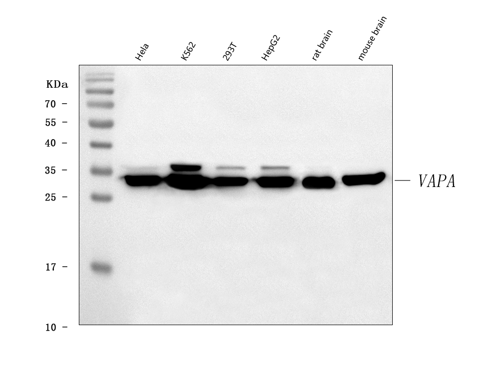
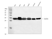
Western blot analysis of VAPA using anti-VAPA antibody. The sample well of each lane was loaded with 30 ug of sample under reducing conditions.IHC
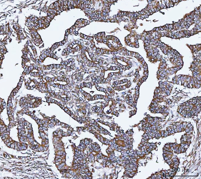
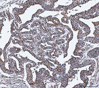
IHC analysis of VAPA using anti-VAPA antibody.IHC
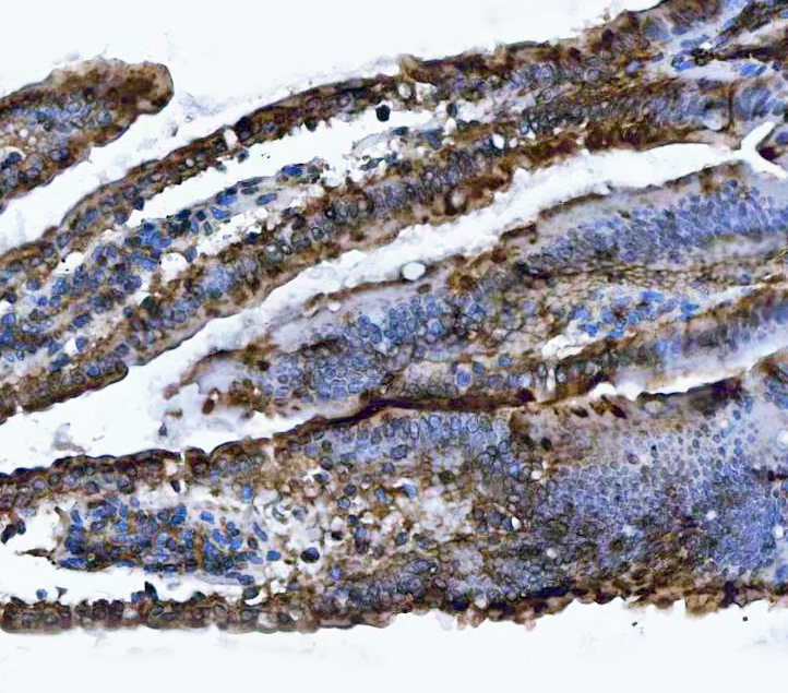
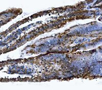
IHC analysis of VAPA using anti-VAPA antibody.IHC
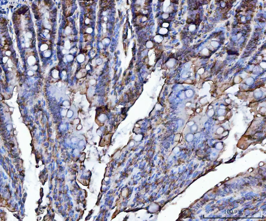
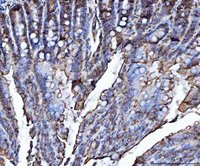
IHC analysis of VAPA using anti-VAPA antibody.ICC/IF
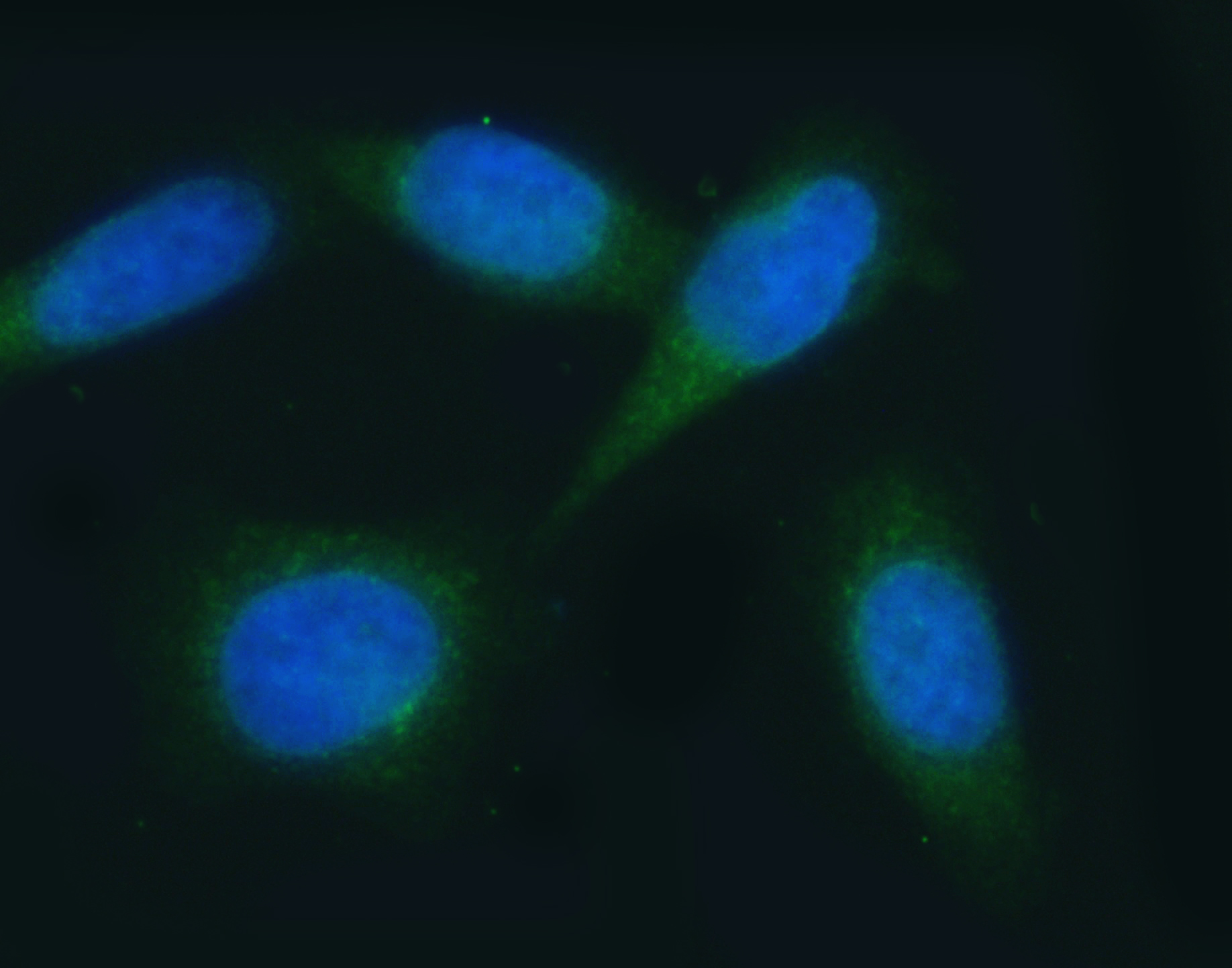
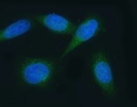
ICC/IF analysis of VAPA using anti-VAPA antibody.IF-P
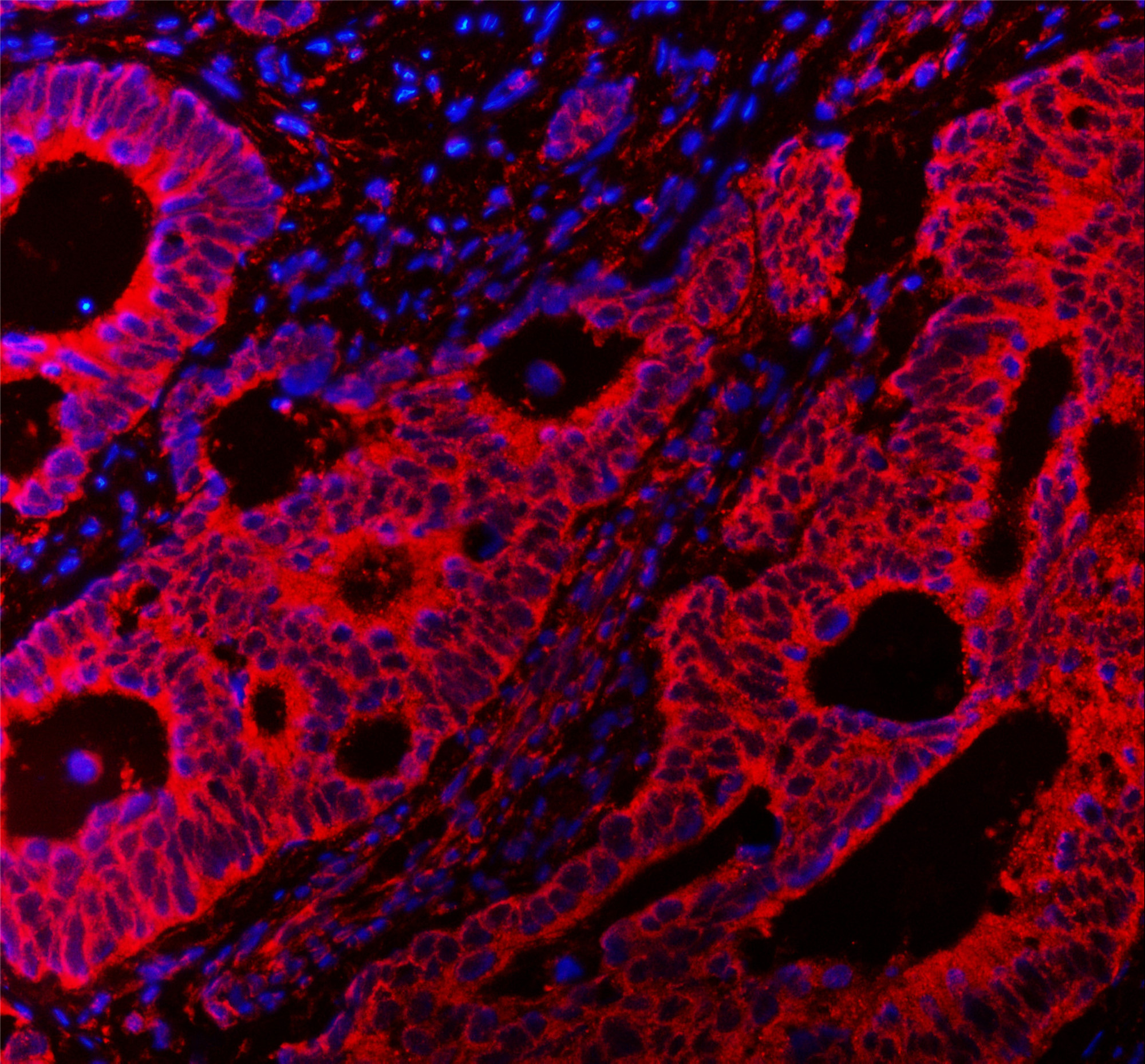
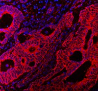
IF analysis using anti- VAPA antibody detected in paraffin-embedded section of human colorectal adenocarcinoma tissue. The tissue section were stained using the cy3-conjugated Anti-rabbit IgG Secondary Antibody (red) and counterstained with DAPI (blue).IF-P
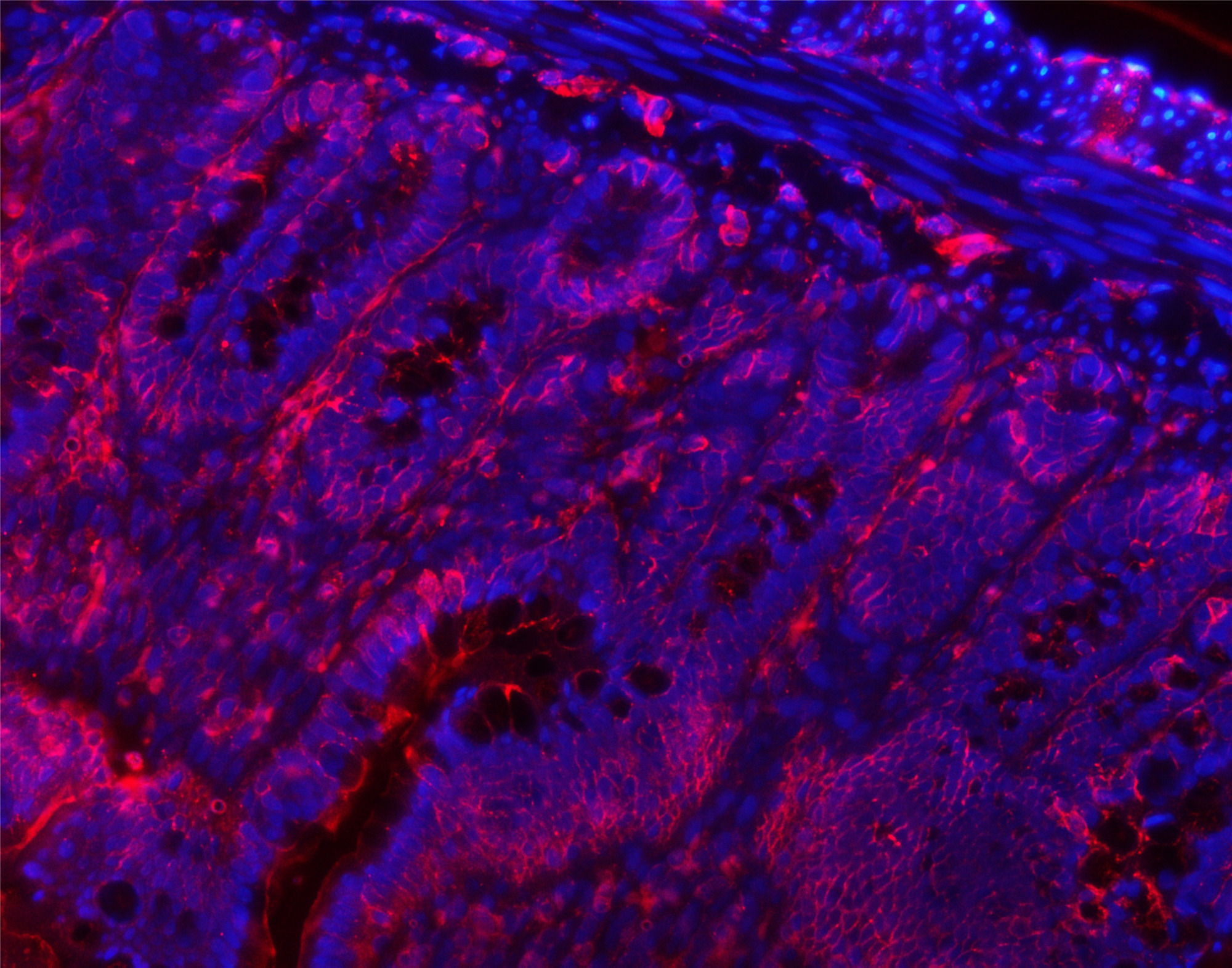
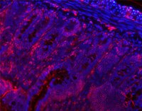
IF analysis using anti- VAPA antibody detected in paraffin-embedded section of rat colon tissue. The tissue section were stained using the cy3-conjugated Anti-rabbit IgG Secondary Antibody (red) and counterstained with DAPI (blue).FC
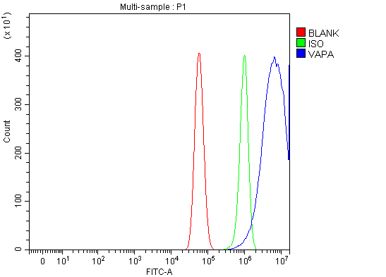
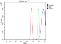
Flow Cytometry analysis of U87 cells using anti-VAPA antibody.| 抗体类型 | Primary Antibodies |
|---|---|
| 免疫原 | A synthetic peptide corresponding to a sequence at the N-terminus of human VAPA, which shares 94.1% amino acid (aa) sequence identity with mouse and rat VAPA. |
| 克隆性 | Polyclonal |
| 同种型 | IgG |
| 宿主 | Rabbit |
| 已测试的应用 | FCICC/IFIF-PIHCWB |
| 物种反应性 | HumanMouseRat |
| 浓度 | 0.5mg/ml |
| 纯化 | Affinity purified |
| 基因名称 | VAPA |
| 基因别名 | VAP-A; VAP33; VAMP-A; VAP-33; hVAP-33 |
| 基因全称 | VAMP associated protein A |
| 基因简介 | The protein encoded by this gene is a type IV membrane protein. It is present in the plasma membrane and intracellular vesicles. It may also be associated with the cytoskeleton. This protein may function in vesicle trafficking, membrane fusion, protein complex assembly and cell motility. Alternative splicing occurs at this locus and two transcript variants encoding distinct isoforms have been identified. [provided by RefSeq, Jul 2008] |
| 蛋白别名 | 33 kDa VAMP associated protein; hVAP 33; VAMP A; VAMP associated protein A; VAP 33; VAP A; VAP33; VAPA |
| 分子量 | 28 kDa(Observed band size: 33 kDa) |
| 应用稀释比 |
WB:1:500-1:2000 IHC:1:50-1:400 ICC/IF:1:50-1:400 IF-P:1:50-1:400 FC:1:50-1:200 |
| 亚细胞定位 | Cell junction,Cell membrane,Endoplasmic reticulum,Membrane,Nucleus,Tight junction. |
| 产品形态 | Liquid |
| 缓冲液 | 500 ug/ml antibody with PBS, 0.02% NaN3, 1 mg/ml BSA and 50% glycerol. |
| 保存条件 | 12 months from date of receipt,-20℃ as supplied. |
请在以下方框中输入您的动物实验信息后点击计算,可以得到母液配置方法和体内配方的制备方法:
比如您的给药剂量是10 mg/kg,每只动物体重20 g,给药体积100 μL, 一共给药动物10只,您使用的配方为 10% DMSO + 40% PEG300 + 5% Tween 80 + 45% Saline / PBS / ddH2O, 那么您的工作液浓度为2 mg/mL。
母液配置方法:2 mg 药物溶于 100 μL DMSO ( 母液浓度为 20 mg/mL ), 如您需要配置的浓度超过该产品的溶解度,请先与我们联系。
体内配方的制备方法: 取 100 μL DMSO 母液, 添加 400 μL PEG300 混匀澄清, 再加 50 μL Tween 80, 混匀澄清, 再加 450 μL Saline / PBS / ddH2O 混匀澄清
以上为“体内实验配液计算器”的使用方法举例,并不是具体某个化合物的推荐配制方式,请根据您的实验动物和给药方式选择适当的溶解方案。
方案所需的各类助溶剂如: DMSO, PEG300, PEG400, Tween 80, SBE-β-CD, 玉米油等, 均可在诺渊网站点击购买。
计算结果:
工作液浓度: mg/ml;
DMSO母液配制方法: mg 药物溶于 μL DMSO溶液(母液浓度 mg/mL,
体内配方配制方法:取 μL DMSO母液,加入 μL PEG300,混匀澄清后加入μL Tween 80,混匀澄清后加入 μL Saline/PBS/ddH2O,混匀澄清。
1. 首先保证母液是澄清的;
2.
一定要按照顺序依次将溶剂加入,进行下一步操作之前必须保证上一步操作得到的是澄清的溶液,可采用涡旋、超声或水浴加热等物理方法助溶。


