
WB
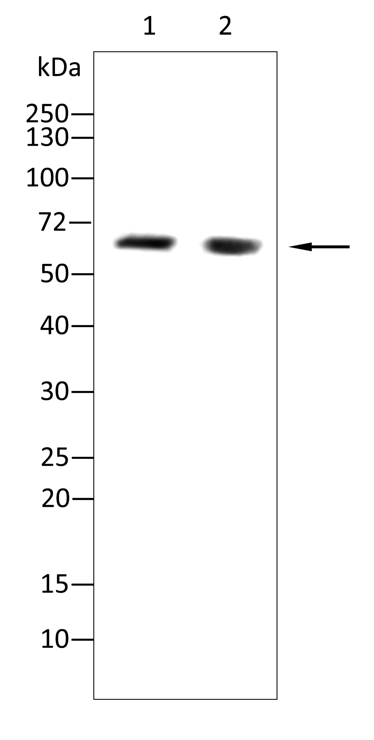
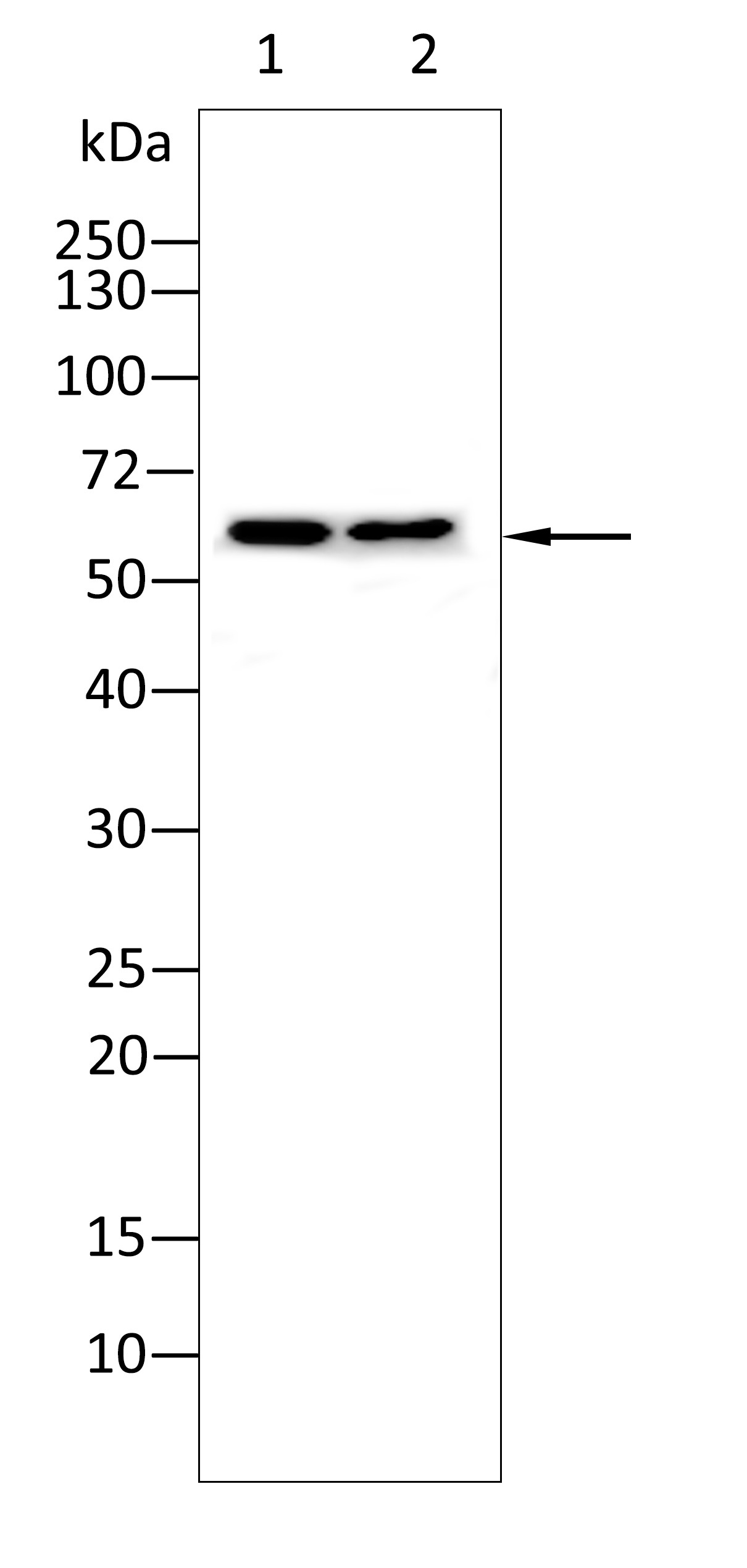
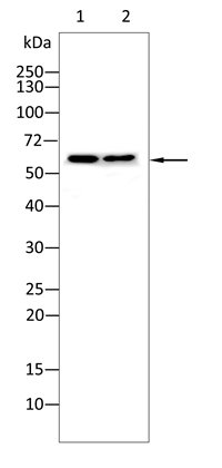
Western blot analysis using PPAR-gamma antibody against A549 (1), Caco-2 (2) cell lysate. 12% SDS-PAGE gel. Sample loading: 20μg /lane. Transfer the proteins onto a PVDF membrane, and block it with TBST plus skimmed milk powder for one hour. Dilute the primary antibody with the antibody diluent at a ratio of 1:1000, and incubate it overnight at 4°C. Wash the membrane three times with TBST, 5 minutes each time. At room temperature, dilute the secondary antibody, Goat Anti-Rabbit IgG(H&L)-HRP, at a ratio of 1:20000 and incubate for one hour. Wash the membrane three times with TBST again, 5 minutes each time. Use ECL for luminescence. staining time: 60S.IHC
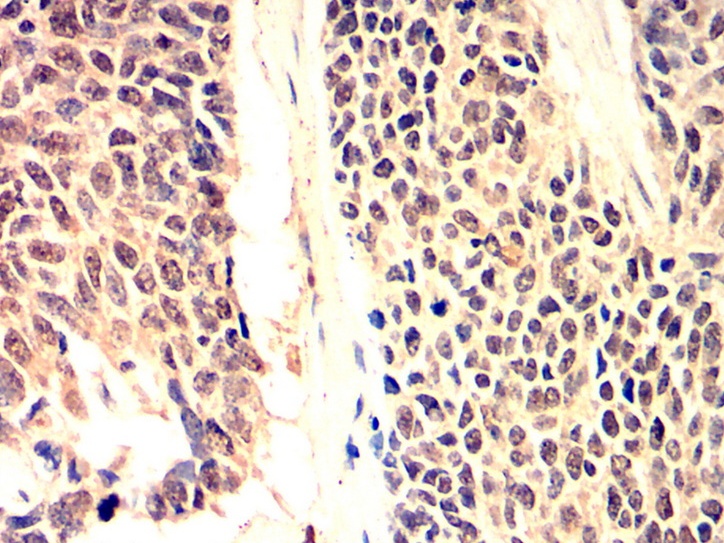
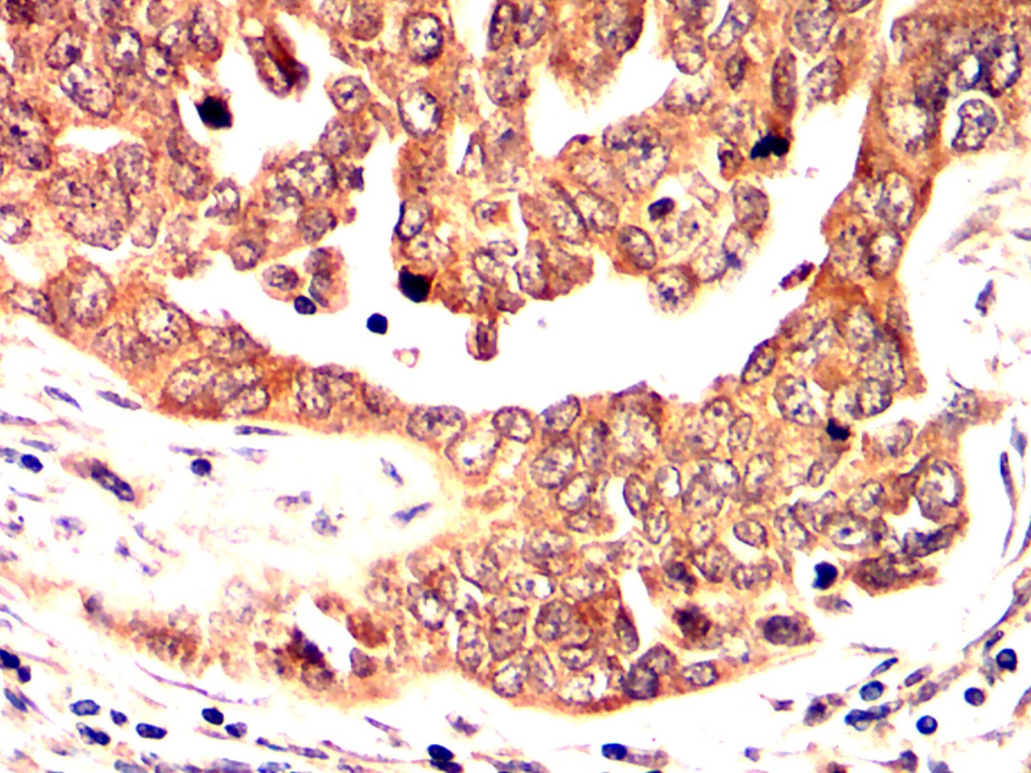
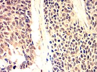
Immunohistochemical analysis of paraffin-embedded Human lung cancer tissues using PPAR-gamma antibody with DAB staining. Pre-treat the sections with heat-mediated antigen retrieval using sodium citrate buffer (pH 6. 0) for 2 minutes. Wash the sections with ddH₂O and PBS. Block the tissue with 10% non-immune goat serum at room temperature for 30 minutes. Incubate the tissue with the primary antibody diluted at a ratio of 1:1500 at 4°C overnight. At room temperature, dilute the secondary antibody, Goat Anti-Rabbit IgG(H&L)-HRP, at a ratio of 1:200 and incubate for one hour. Use DAB as the chromogenic agent. Counterstain the tissue with hematoxylin, and mount the tissue sections with neutral gum.IHC
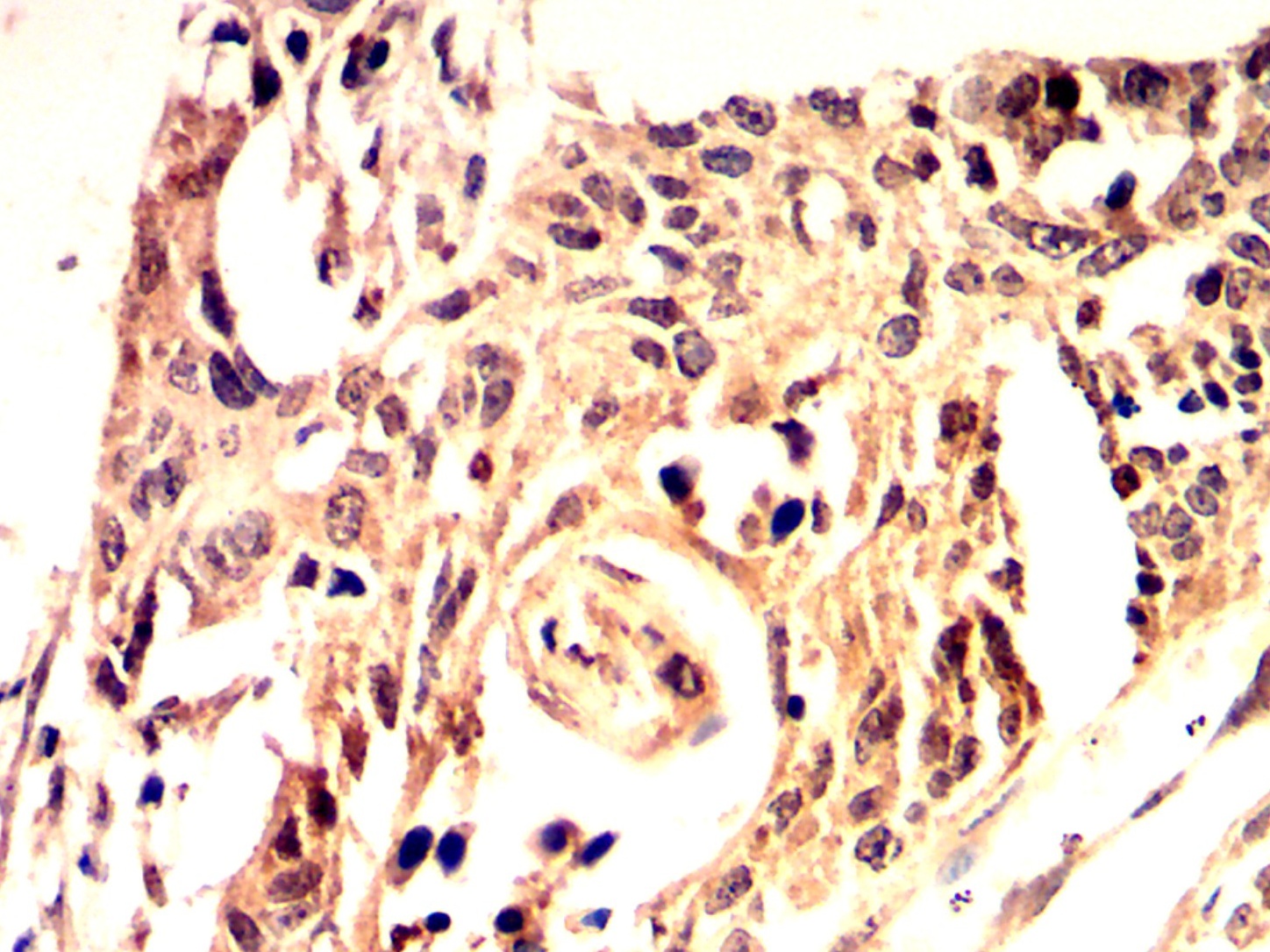
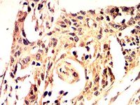
Immunohistochemical analysis of paraffin-embedded Human esophageal cancer tissues using PPAR-gamma antibody with DAB staining. Pre-treat the sections with heat-mediated antigen retrieval using sodium citrate buffer (pH 6. 0) for 2 minutes. Wash the sections with ddH₂O and PBS. Block the tissue with 10% non-immune goat serum at room temperature for 30 minutes. Incubate the tissue with the primary antibody diluted at a ratio of 1:1500 at 4°C overnight. At room temperature, dilute the secondary antibody, Goat Anti-Rabbit IgG(H&L)-HRP, at a ratio of 1:200 and incubate for one hour. Use DAB as the chromogenic agent. Counterstain the tissue with hematoxylin, and mount the tissue sections with neutral gum.IHC
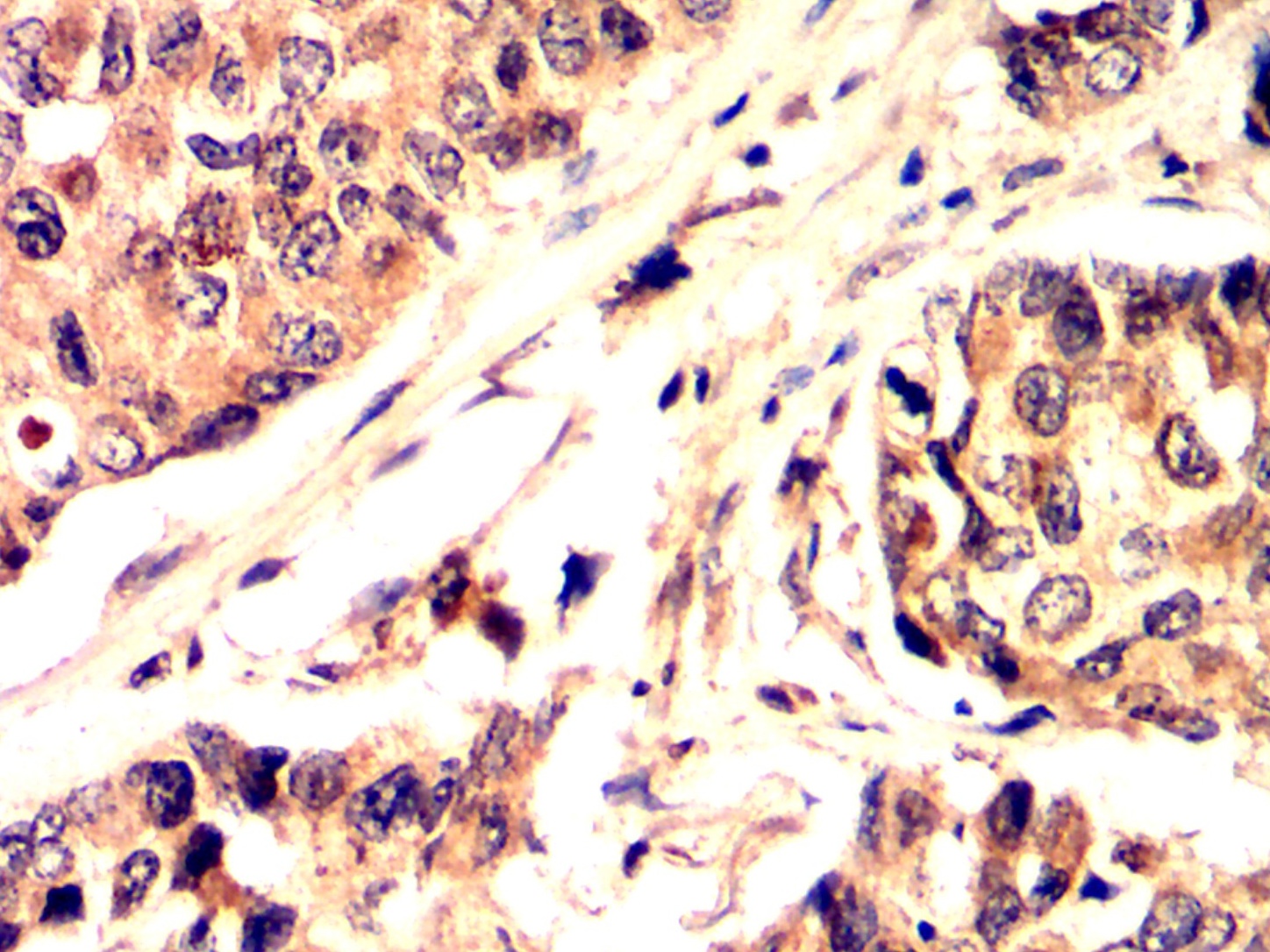
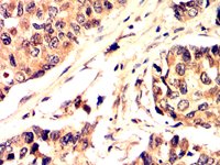
Immunohistochemical analysis of paraffin-embedded Human breast cancer tissues using PPAR-gamma antibody with DAB staining. Pre-treat the sections with heat-mediated antigen retrieval using sodium citrate buffer (pH 6. 0) for 2 minutes. Wash the sections with ddH₂O and PBS. Block the tissue with 10% non-immune goat serum at room temperature for 30 minutes. Incubate the tissue with the primary antibody diluted at a ratio of 1:1500 at 4°C overnight. At room temperature, dilute the secondary antibody, Goat Anti-Rabbit IgG(H&L)-HRP, at a ratio of 1:200 and incubate for one hour. Use DAB as the chromogenic agent. Counterstain the tissue with hematoxylin, and mount the tissue sections with neutral gum.IHC
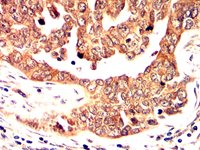
Immunohistochemical analysis of paraffin-embedded Human ovarian cancer tissues using PPAR-gamma antibody with DAB staining. Pre-treat the sections with heat-mediated antigen retrieval using sodium citrate buffer (pH 6. 0) for 2 minutes. Wash the sections with ddH₂O and PBS. Block the tissue with 10% non-immune goat serum at room temperature for 30 minutes. Incubate the tissue with the primary antibody diluted at a ratio of 1:1500 at 4°C overnight. At room temperature, dilute the secondary antibody, Goat Anti-Rabbit IgG(H&L)-HRP, at a ratio of 1:200 and incubate for one hour. Use DAB as the chromogenic agent. Counterstain the tissue with hematoxylin, and mount the tissue sections with neutral gum.WB
Western blot analysis using PPAR-gamma antibody against Mouse intestine(1), Rat stomach(2) tissue. 12% SDS-PAGE gel. Sample loading: 20μg /lane. Transfer the proteins onto a PVDF membrane, and block it with TBST plus skimmed milk powder for one hour. Dilute the primary antibody with the antibody diluent at a ratio of 1:1000-1:2000, and incubate it overnight at 4°C. Wash the membrane three times with TBST, 5 minutes each time. At room temperature, dilute the secondary antibody, Goat Anti-Rabbit IgG(H&L)-HRP, at a ratio of 1:20000 and incubate for one hour. Wash the membrane three times with TBST again, 5 minutes each time. Use ECL for luminescence. staining time: 60S.ELISA
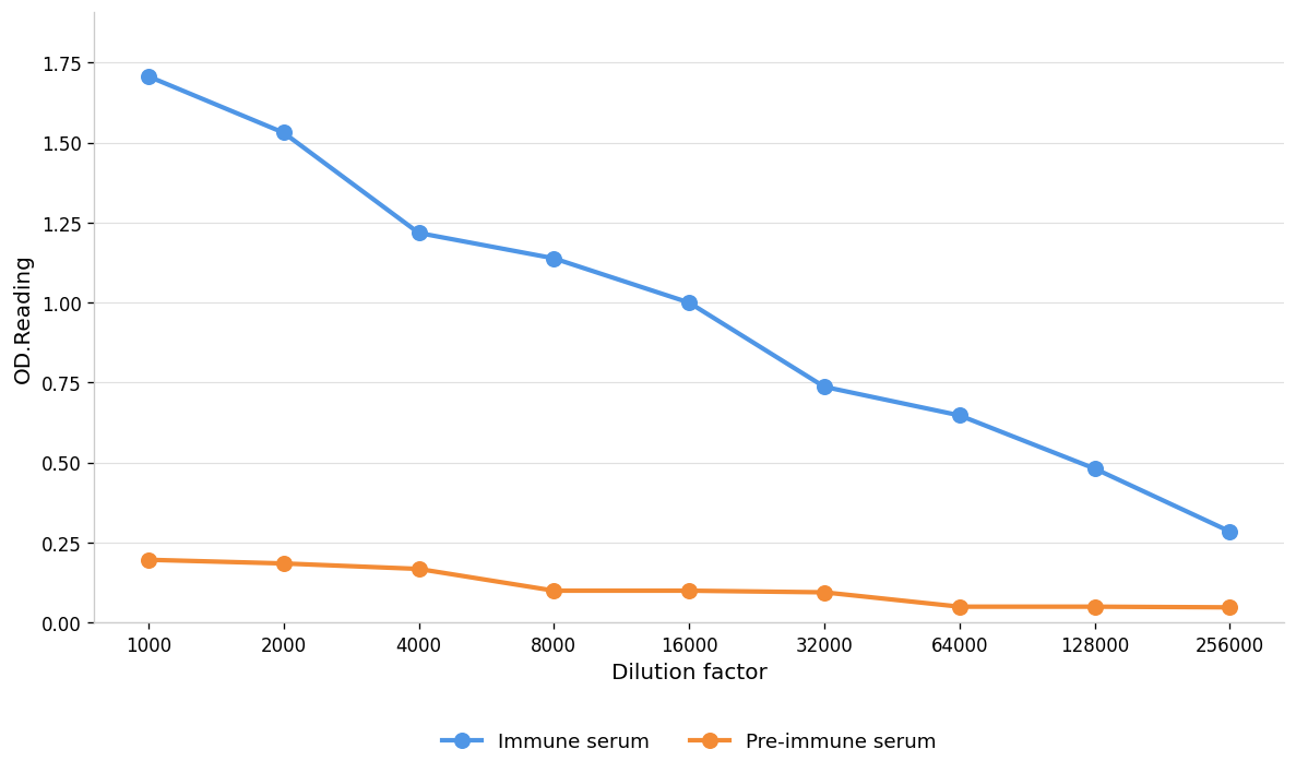
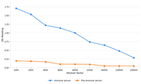
Coat the plate with immunizing antigen. Blue line:Immune serum; Orange line: Pre-immune serum.| 抗体类型 | Primary Antibodies |
|---|---|
| 免疫原 | Polypeptide |
| 克隆性 | Polyclonal |
| 同种型 | IgG |
| 宿主 | Rabbit |
| 来源 | Rabbit |
| 已测试的应用 | ELISAIHCWB |
| 物种反应性 | HumanMouseRat |
| 浓度 | 1mg/ml |
| 纯化 | Protein A |
| 基因名称 | PPARG |
| 基因别名 | CIMT1; FPLD3; GLM1; NR1C3; PPAR-GAMMA; PPAR-gamma2; PPARG1; PPARG2; PPARG5; PPARGAMMA; PPARgamma2 |
| 基因全称 | peroxisome proliferator activated receptor gamma |
| 基因简介 | This gene encodes a member of the peroxisome proliferator-activated receptor (PPAR) subfamily of nuclear receptors. PPARs form heterodimers with retinoid X receptors (RXRs) and these heterodimers regulate transcription of various genes. Three subtypes of PPARs are known: PPAR-alpha, PPAR-delta, and PPAR-gamma. The protein encoded by this gene is PPAR-gamma and is a regulator of adipocyte differentiation. Additionally, PPAR-gamma has been implicated in the pathology of numerous diseases including obesity, diabetes, atherosclerosis and cancer. Alternatively spliced transcript variants that encode different isoforms have been described. [provided by RefSeq, Jul 2008] |
| 蛋白别名 | HUMPPARG; ligand-activated nuclear transcription factor; ligand-activated transcription factor; nuclear hormone receptor; nuclear hormone receptor; PPARG; nuclear receptor; nuclear receptor family member; Pparg; Nuclear receptor subfamily 1 group C member 3; nuclear receptor transcription factor; OTTHUMP00000185032; OTTHUMP00000185036; peroxisome proliferative activated receptor, gamma; peroxisome proliferator; peroxisome proliferator activated receptor; peroxisome proliferator activated receptor gamma; peroxisome proliferator activated receptor gamma 2; peroxisome proliferator activated receptor gamma 4; peroxisome proliferator activated receptor gamma-1; peroxisome proliferator activatived receptor gamma; peroxisome proliferator activator receptor, gamma; peroxisome proliferator-activated receptor gamma 1; peroxisome proliferator-activated receptor gamma 1-a; peroxisome proliferator-activated receptor gamma 1-b; peroxisome proliferator-activated receptor gamma 2; peroxisome proliferator-activated receptor-gamma 5; peroxisome proliferator-activated receptor-gamma splicing; PPAR gamma; PPAR gamma 1; PPAR gamma 3; PPAR-gamma; PPAR-gamma-1a; PPAR-gamma-1b; Pparg; PPARgamma; PPARy; transcription factor |
| 分子量 | 58 kDa |
| Uniprot ID | P37231 |
| 应用稀释比 |
WB:1:1000-1:2000 IHC:1:200-1:2000 ELISA:1:1000-1:10000 |
| 修饰 | Unmodification |
| 标记 | Unconjugated |
| 亚细胞定位 | Nucleus |
| 产品形态 | Liquid |
| 缓冲液 | Purified antibody in PBS with 0.05% sodium azi,40% Glycerol. |
| 保存条件 | Shipped at 4°C. Store at +4°C short term (1-2 weeks). Store at -20°C long term. Avoid freeze / thaw cycle. |
请在以下方框中输入您的动物实验信息后点击计算,可以得到母液配置方法和体内配方的制备方法:
比如您的给药剂量是10 mg/kg,每只动物体重20 g,给药体积100 μL, 一共给药动物10只,您使用的配方为 10% DMSO + 40% PEG300 + 5% Tween 80 + 45% Saline / PBS / ddH2O, 那么您的工作液浓度为2 mg/mL。
母液配置方法:2 mg 药物溶于 100 μL DMSO ( 母液浓度为 20 mg/mL ), 如您需要配置的浓度超过该产品的溶解度,请先与我们联系。
体内配方的制备方法: 取 100 μL DMSO 母液, 添加 400 μL PEG300 混匀澄清, 再加 50 μL Tween 80, 混匀澄清, 再加 450 μL Saline / PBS / ddH2O 混匀澄清
以上为“体内实验配液计算器”的使用方法举例,并不是具体某个化合物的推荐配制方式,请根据您的实验动物和给药方式选择适当的溶解方案。
方案所需的各类助溶剂如: DMSO, PEG300, PEG400, Tween 80, SBE-β-CD, 玉米油等, 均可在诺渊网站点击购买。
计算结果:
工作液浓度: mg/ml;
DMSO母液配制方法: mg 药物溶于 μL DMSO溶液(母液浓度 mg/mL,
体内配方配制方法:取 μL DMSO母液,加入 μL PEG300,混匀澄清后加入μL Tween 80,混匀澄清后加入 μL Saline/PBS/ddH2O,混匀澄清。
1. 首先保证母液是澄清的;
2.
一定要按照顺序依次将溶剂加入,进行下一步操作之前必须保证上一步操作得到的是澄清的溶液,可采用涡旋、超声或水浴加热等物理方法助溶。


