WB
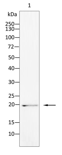
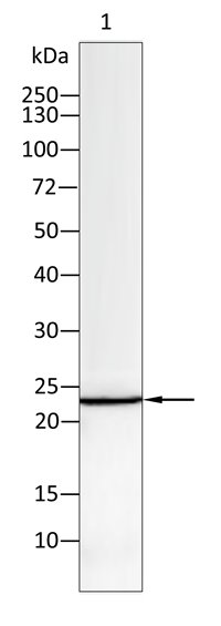
Western blot analysis using p21 Cip1 antibody against HepG2 (1) cell lysate. 12% SDS-PAGE gel. Sample loading: 20μg /lane. Transfer the proteins onto a PVDF membrane, and block it with TBST plus skimmed milk powder for one hour. Dilute the primary antibody with the antibody diluent at a ratio of 1:1000, and incubate it overnight at 4°C. Wash the membrane three times with TBST, 5 minutes each time. At room temperature, dilute the secondary antibody, Goat Anti-Rabbit IgG(H&L)-HRP, at a ratio of 1:20000 and incubate for one hour. Wash the membrane three times with TBST again, 5 minutes each time. Use ECL for luminescence. staining time: 60S.IHC
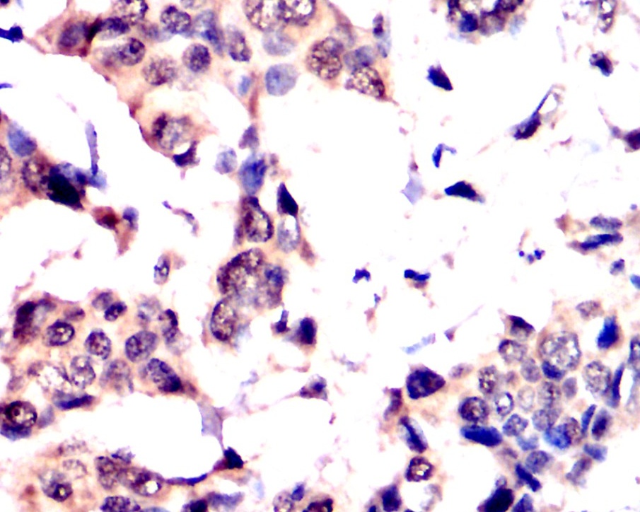
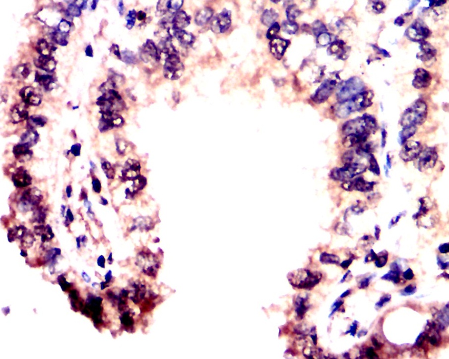
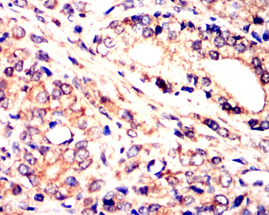
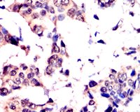
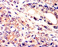
Immunohistochemical analysis of paraffin-embedded Human breast cancer tissues using p21 Cip1 antibody with DAB staining. Pre-treat the sections with heat-mediated antigen retrieval using sodium citrate buffer (pH 6. 0) for 2 minutes. Wash the sections with ddH₂O and PBS. Block the tissue with 10% non-immune goat serum at room temperature for 30 minutes. Incubate the tissue with the primary antibody diluted at a ratio of 1:1500 at 4°C overnight. At room temperature, dilute the secondary antibody, Goat Anti-Rabbit IgG(H&L)-HRP, at a ratio of 1:200 and incubate for one hour. Use DAB as the chromogenic agent. Counterstain the tissue with hematoxylin, and mount the tissue sections with neutral gum.ICC/IF
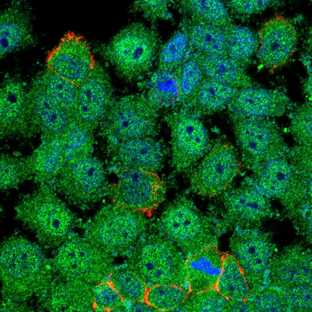
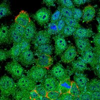
Immunofluorescence analysis of L-02 cells using p21 Cip1 antibody (green). Blue: DAPI fluorescent DNA dye. Red: Actin filaments have been labeled withTSZ® 594-Phalloidin. Cells are fixed in 4% paraformaldehyde at room temperature for 20 minutes. Then, they are permeabilized with a PBS solution containing 0. 1% Triton X-100 at room temperature for 15 minutes. Subsequently, the cells are blocked with 10% non - immune goat serum at room temperature for 1 hour. The cells are incubated overnight at 4°C with the primary antibody diluted 1:100 in PBS. The secondary antibody, TSZ® 488 Goat Ant-Rabbit IgG(H&L) (Green) is diluted at a ratio of 1:400 and incubated with the cells for 1 hour. Nuclear DNA is labeled with DAPI (Blue). F-actin is stained with TSZ® 594-Phalloidin (Red), diluted 1:100 for 30 minutes.IHC
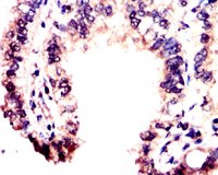
Immunohistochemical analysis of paraffin-embedded Human lung cancer tissues using p21 Cip1 antibody with DAB staining. Pre-treat the sections with heat-mediated antigen retrieval using sodium citrate buffer (pH 6. 0) for 2 minutes. Wash the sections with ddH₂O and PBS. Block the tissue with 10% non-immune goat serum at room temperature for 30 minutes. Incubate the tissue with the primary antibody diluted at a ratio of 1:1500 at 4°C overnight. At room temperature, dilute the secondary antibody, Goat Anti-Rabbit IgG(H&L)-HRP, at a ratio of 1:200 and incubate for one hour. Use DAB as the chromogenic agent. Counterstain the tissue with hematoxylin, and mount the tissue sections with neutral gum.IHC
Immunohistochemical analysis of paraffin-embedded Human prostate cancer tissues using p21 Cip1 antibody with DAB staining. Pre-treat the sections with heat-mediated antigen retrieval using sodium citrate buffer (pH 6. 0) for 2 minutes. Wash the sections with ddH₂O and PBS. Block the tissue with 10% non-immune goat serum at room temperature for 30 minutes. Incubate the tissue with the primary antibody diluted at a ratio of 1:1500 at 4°C overnight. At room temperature, dilute the secondary antibody, Goat Anti-Rabbit IgG(H&L)-HRP, at a ratio of 1:200 and incubate for one hour. Use DAB as the chromogenic agent. Counterstain the tissue with hematoxylin, and mount the tissue sections with neutral gum.WB
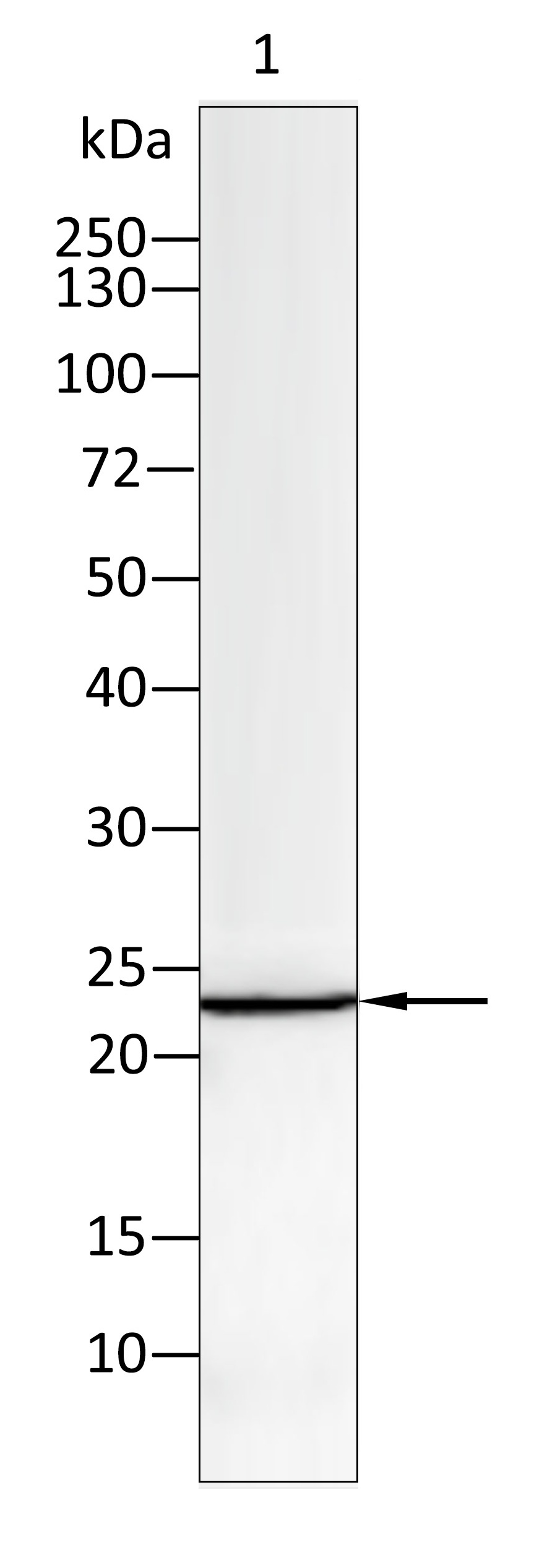
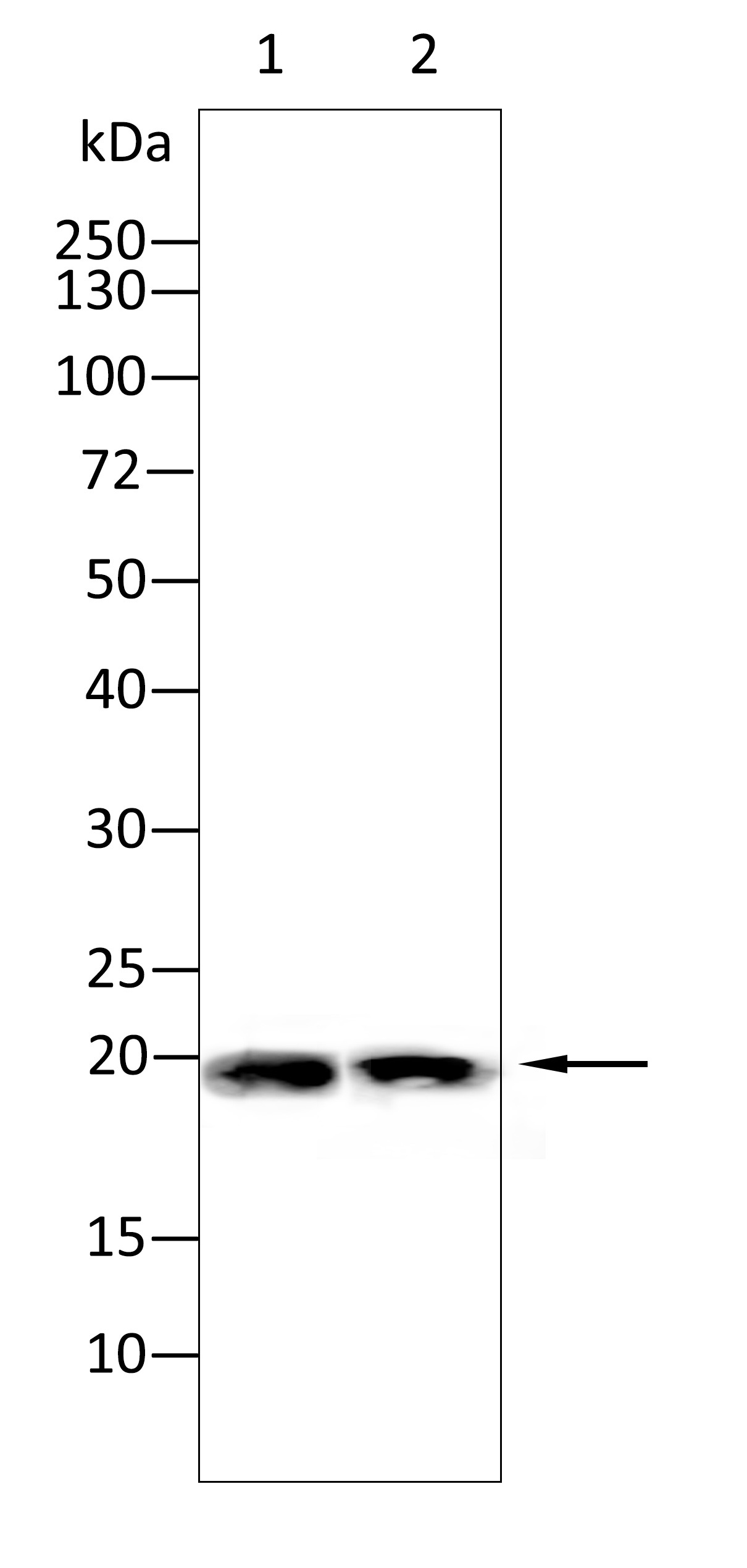
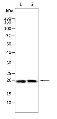
Western blot analysis using p21 Cip1 antibody against Mouse brain(1), Mouse testes(2) tissue. 12% SDS-PAGE gel. Sample loading: 20μg /lane. Transfer the proteins onto a PVDF membrane, and block it with TBST plus skimmed milk powder for one hour. Dilute the primary antibody with the antibody diluent at a ratio of 1:1000-1:2000, and incubate it overnight at 4°C. Wash the membrane three times with TBST, 5 minutes each time. At room temperature, dilute the secondary antibody, Goat Anti-Rabbit IgG(H&L)-HRP, at a ratio of 1:20000 and incubate for one hour. Wash the membrane three times with TBST again, 5 minutes each time. Use ECL for luminescence. staining time: 60S.ELISA
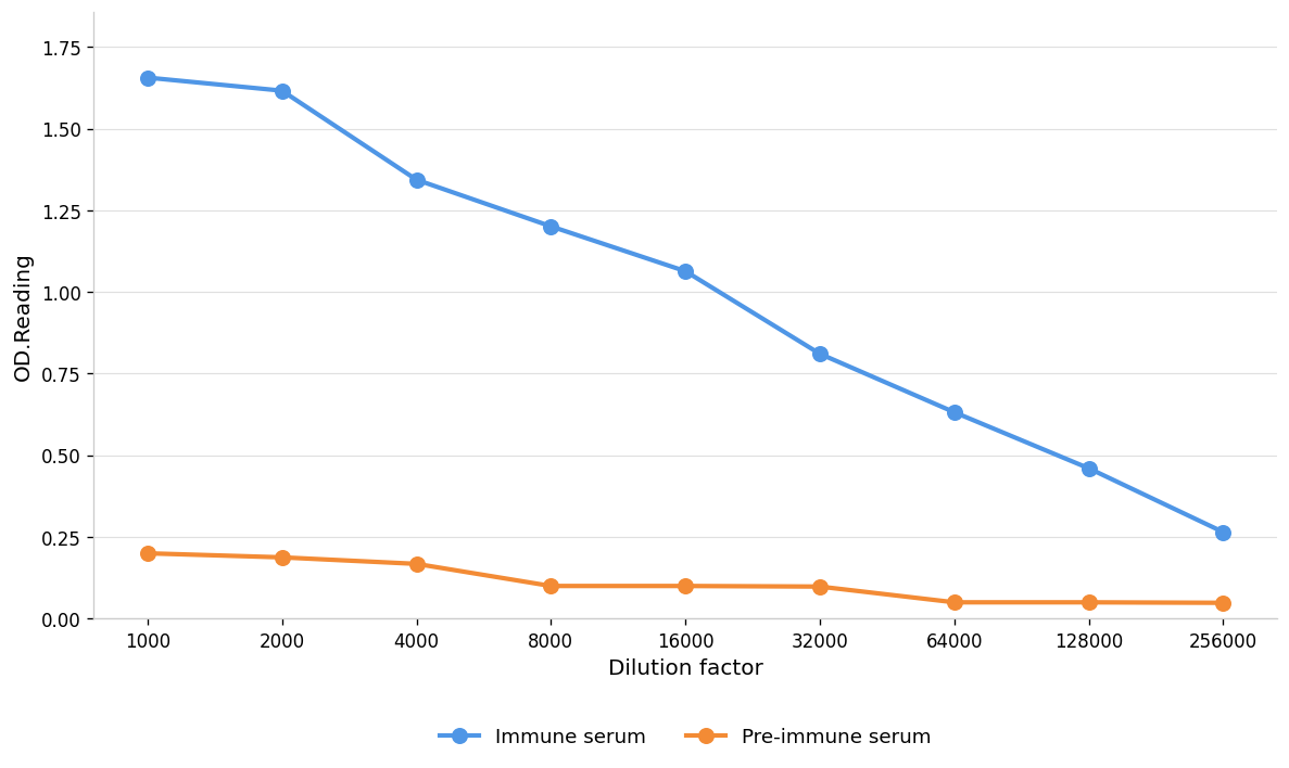
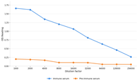
Coat the plate with immunizing antigen. Blue line:Immune serum; Orange line: Pre-immune serum.| 抗体类型 | Primary Antibodies |
|---|---|
| 免疫原 | Polypeptide |
| 克隆性 | Polyclonal |
| 同种型 | IgG |
| 宿主 | Rabbit |
| 已测试的应用 | ELISAICC/IFIHCWB |
| 物种反应性 | HumanMouseRat |
| 浓度 | 1mg/ml |
| 纯化 | Protein A |
| 基因名称 | CDKN1A |
| 基因别名 | CAP20; CDKI; CDKN1; CIP1; MDA-6; mda6; P21; p21CIP1; p21WAF; SDI1; WAF1 |
| 基因全称 | cyclin dependent kinase inhibitor 1A |
| 基因简介 | This gene encodes a potent cyclin-dependent kinase inhibitor. The encoded protein binds to and inhibits the activity of cyclin-cyclin-dependent kinase2 or -cyclin-dependent kinase4 complexes, and thus functions as a regulator of cell cycle progression at G1. The expression of this gene is tightly controlled by the tumor suppressor protein p53, through which this protein mediates the p53-dependent cell cycle G1 phase arrest in response to a variety of stress stimuli. This protein can interact with proliferating cell nuclear antigen, a DNA polymerase accessory factor, and plays a regulatory role in S phase DNA replication and DNA damage repair. This protein was reported to be specifically cleaved by CASP3-like caspases, which thus leads to a dramatic activation of cyclin-dependent kinase2, and may be instrumental in the execution of apoptosis following caspase activation. Mice that lack this gene have the ability to regenerate damaged or missing tissue. Multiple alternatively spliced variants have been found for this gene. [provided by RefSeq, Sep 2015] |
| 蛋白别名 | Activating Fragment 1; alternate gene name=WAF1; CDK-interacting protein 1; CDK-interaction protein 1; CDKN1A; CDNK1A; Cip1); cyclin kinase inhibitor; Cyclin-dependent kinase inhibitor 1A (p21; cyclin-dependent kinase inhibitor 1A (p21, Cip1); DNA synthesis inhibitor; melanoma differentiation associated protein 6; melanoma differentiation-associated protein; OTTHUMP00000016298; p21Cip1/Waf1; putative DNA synthesis inhibitor; SLC12A9; Wild type p53 activated fragment 1 (WAF1); wild type p53 activated fragment-1; wild-type p53-activated fragment 1 |
| 分子量 | 18 kDa |
| Uniprot ID | P38936 |
| 应用稀释比 |
WB:1:1000-1:2000 IHC:1:200-1:2000 ICC/IF:1:100-1:500 ELISA:1:1000-1:10000 |
| 修饰 | Unmodification |
| 标记 | Unconjugated |
| 亚细胞定位 | Cytoplasm. Nucleus |
| 产品形态 | Liquid |
| 缓冲液 | Purified antibody in PBS with 0.05% PC300,40% Glycerol. |
| 保存条件 | Shipped at 4°C. Store at +4°C short term (1-2 weeks). Store at -20°C long term. Avoid freeze / thaw cycle. |
请在以下方框中输入您的动物实验信息后点击计算,可以得到母液配置方法和体内配方的制备方法:
比如您的给药剂量是10 mg/kg,每只动物体重20 g,给药体积100 μL, 一共给药动物10只,您使用的配方为 10% DMSO + 40% PEG300 + 5% Tween 80 + 45% Saline / PBS / ddH2O, 那么您的工作液浓度为2 mg/mL。
母液配置方法:2 mg 药物溶于 100 μL DMSO ( 母液浓度为 20 mg/mL ), 如您需要配置的浓度超过该产品的溶解度,请先与我们联系。
体内配方的制备方法: 取 100 μL DMSO 母液, 添加 400 μL PEG300 混匀澄清, 再加 50 μL Tween 80, 混匀澄清, 再加 450 μL Saline / PBS / ddH2O 混匀澄清
以上为“体内实验配液计算器”的使用方法举例,并不是具体某个化合物的推荐配制方式,请根据您的实验动物和给药方式选择适当的溶解方案。
方案所需的各类助溶剂如: DMSO, PEG300, PEG400, Tween 80, SBE-β-CD, 玉米油等, 均可在诺渊网站点击购买。
计算结果:
工作液浓度: mg/ml;
DMSO母液配制方法: mg 药物溶于 μL DMSO溶液(母液浓度 mg/mL,
体内配方配制方法:取 μL DMSO母液,加入 μL PEG300,混匀澄清后加入μL Tween 80,混匀澄清后加入 μL Saline/PBS/ddH2O,混匀澄清。
1. 首先保证母液是澄清的;
2.
一定要按照顺序依次将溶剂加入,进行下一步操作之前必须保证上一步操作得到的是澄清的溶液,可采用涡旋、超声或水浴加热等物理方法助溶。